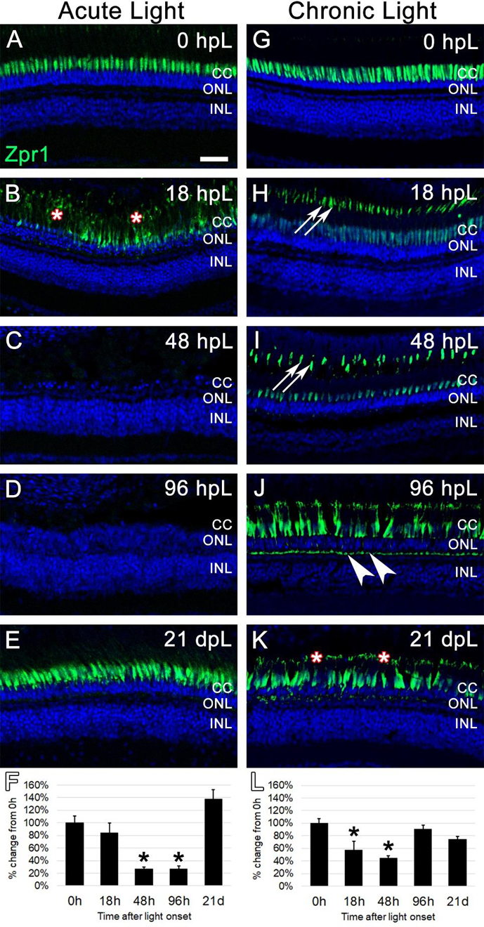
Figure 6. Analysis of cone photoreceptor degeneration/regeneration during acute and chronic light treatments.

In undamaged retinas, Zpr1 immunostaining was observed in the perinuclear domain of red/green double cones (A and G). In the acute group (A-E), Zpr1 immunolabeling was observed throughout the cytoplasm of hypertrophied and degenerating cones at 18 hpL (B, asterisks). Nearly all double cone photoreceptors were degenerated and Zpr1 immunolabeling was not detected at 48 hpL and 96 hpL (C, D). At 21 dpL, newly regenerated red/green double cones were observed by the return of Zpr1 immunostaining. Graphic representation of the percent-change in Zpr1 immunostaining from 0 hpL at each time point (F). In the chronic group (G-H), hypertrophy of cone photoreceptors was observed at 18 dpL (H), concomitant with a down-regulation of Zpr1 immunostaining in the perinuclear domain and an upregulation of immunostaining of the cone outer segments (H, arrowheads). This expression pattern persisted through 48 hpL (I). At 96 hpL, cone hypertrophy was readily apparent, with Zpr1 immunolabeling throughout the cone cytoplasm, including the cone pedicles (arrowheads). At 21 dpL, cone photoreceptor hypertrophy persisted, and Zpr1 immunolabeling was suggestive of cone outer segment truncation/degeneration (K, asterisks; compare outer segments in H and I to K). Graphic representation of the percent-change in Zpr1 immunostaining from 0 hpL at each time point (L). hpL: hours post light onset; CC; cone cell nuclei; ONL: outer nuclear layer; INL: inner nuclear layer; asterisks in F and L: significantly different from 0 hpL (p<0.05) as determined by post-hoc Tukey test from one-way ANOVA (N=5–6 retinas per time point). Scale bar=25 microns.
