
Figure 8. Histological sections of photoreceptor degeneration and microglial localization during acute and chronic light treatments.
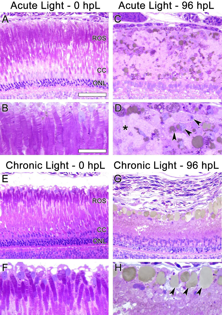
In undamaged retinas (A, B, E, and F) the disc-like structures of ROS were clearly visible. In the acute group at 96 hpL (C, D), rod and cone photoreceptors were completely absent. The resultant cellular debris field (D, asterisk) contained many cells with amoeboid morphology containing small vacuoles/phagosomes (D, arrowheads), likely microglia. In the chronic group at 96 hpL (G, H), ROS and cone outer segments appeared to be absent, but both rod and cone photoreceptor nuclei were still present (G). Near the RPE/ROS interface, cells with amoeboid morphology containing large vacuoles/phagosomes were present (H, arrowheads). Based on location and morphology, these were also likely microglia.