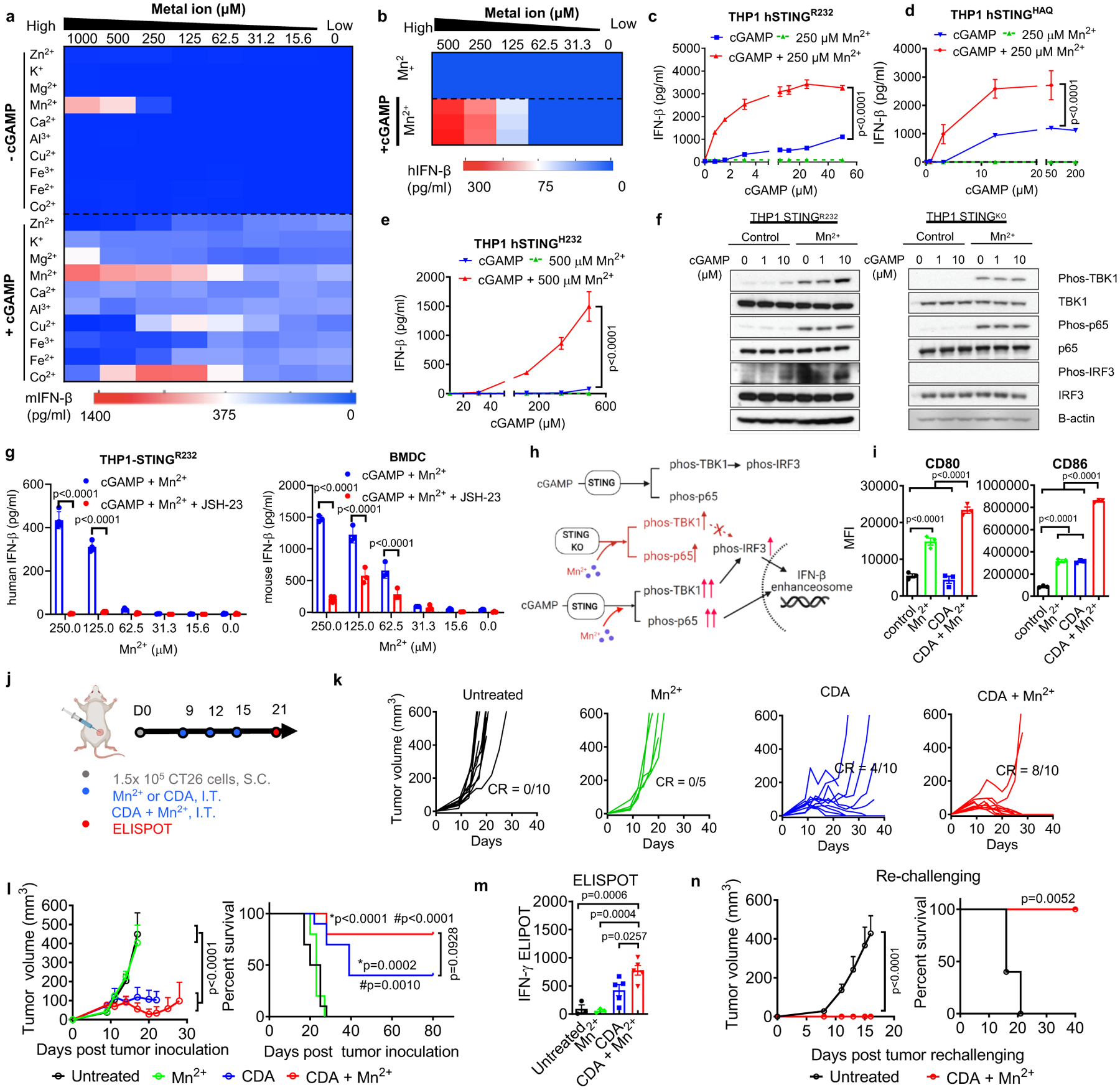
Figure 2. Mn2+ augments type-I IFN activity of STING agonists.

a) BMDCs or b) THP1 cells were incubated with various concentrations of metal ions ± 5 μM cGAMP, and after 24 h, IFN-β secretion was quantified. c–e) THP1 cells expressing c) hSTINGR232, d) hSTINGHAQ, or e) hSTINGH232 were treated for 24 h with cGAMP ± Mn2+, followed by quantification of IFN-β production. f) THP-1 STINGR232 or THP-1 STINGKO cells were incubated with increasing concentrations of cGAMP ± 250 μM Mn2+ for 6 hours, followed by Immunoblotting for marker proteins in the STING-IFN-I pathway. Shown are representative data from two independent experiments with similar results. g) Pharmacological inhibition of p65 nucleus translocation inhibited Mn2+-potentiated IFN-β production. h) Proposed mechanism of Mn2+-mediated potentiation of STING agonist via STING-independent TBK1 and p65 phosphorylation and STING-dependent IRF3 phosphorylation. Activation of p65 and IRF3 further facilitates assembly of the IFN-β transcriptional enhanceosome. i) BMDCs treated with 5 μM CDA, 250 μM Mn2+, or their combination for 24 h were analyzed for activation by flow cytometry. j–n) CT26 tumor-bearing BALB/c mice were treated by I.T. administration with 20 μg CDA, 17.5 μg Mn2+, or their combination on days 9, 12, and 15 (j). Mice were monitored for tumor growth (k, l) and survival (l). m) AH1-specific T-cells among PBMCs were assessed by ELISPOT on day 21. n) Survivors from CDA + Mn2+ group were re-challenged with CT26 cells on day 80. Data represent mean ± SEM, from a representative experiment from 2–3 independent experiments with n = 3–4 (c–e, g, i) and n = 5–10 (k–n) biologically independent samples. Data were analyzed by (i, m) one-way ANOVA or (c–e, g, l, n) two-way ANOVA with Bonferroni’s multiple comparisons test, or (l, n) log-rank (Mantel-Cox) test. *p and #p in (j) denote statistical significance relative to the untreated or Mn2+ group, respectively. Figure 2h and 2j were created using BioRender.com.
