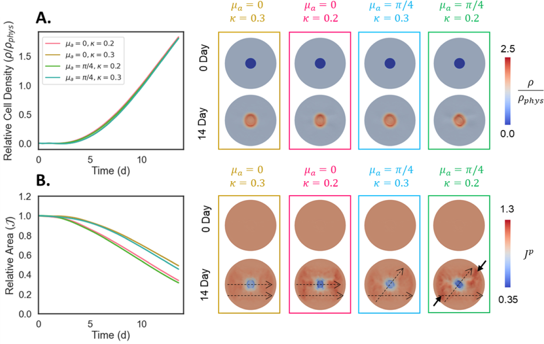
Figure 9:

Effect of dermal scaffold (Oligomer-40) fiber orientation relative to normal surrounding skin (μa) and fiber dispersion (κ) on A. cellularization and B. contraction of wounds. Scaffolds with relatively isotropic fiber distributions (κ = 0.3) yield slower and more uniform circular contraction, while aligned scaffolds (κ = 0.2) contract faster. Scaffold fiber orientation dictates the angle of contraction during wound healing. When scaffold fibers are oriented parallel to the surrounding skin (μa = 0) vertically-oriented scars result, while angled scaffolds (μa = π/4) produce asymmetric contraction fields. Dashed arrows show direction of fibers in the graft and surrounding tissue. Solid arrows emphasize the asymmetric deformation field.
