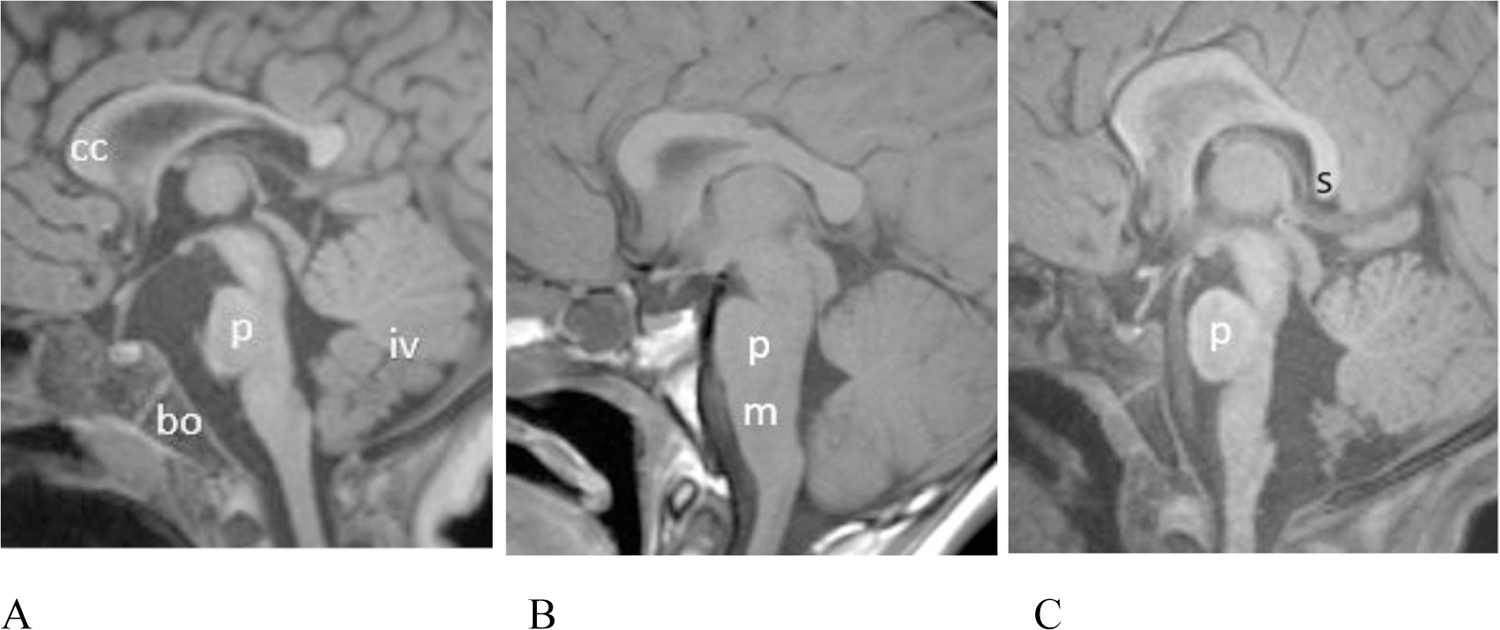
Figure 3.

(A–C) Midline sagittal T1 weighted MR images showing variable pontine dysmorphism. (A) Patient # 17, 15 month old female. T1 MPPRAGE image shows a relative short pons (p) that is mildly narrowed in AP diameter, especially superiorly. There is hypoplasia of the inferior vermis (iv). The corpus callosum (cc) is mildly thinned. Note the normal basiocciput (bo). (B) Patient #6, 11 year old male. Sagittal spin echo T1 weighted image shows that the pons (p) is slightly shortened in height and there is an unusually obtuse angle between the ventral aspect of the pons and medulla (m). (C) Patient #14 (22 month old female). T1 MPRAGE image shows that the pons is mildly shortened in height. The vermis is uplifted. The posterior body of the corpus callosum is mildly thinned and the splenium has a vertical orientation.
