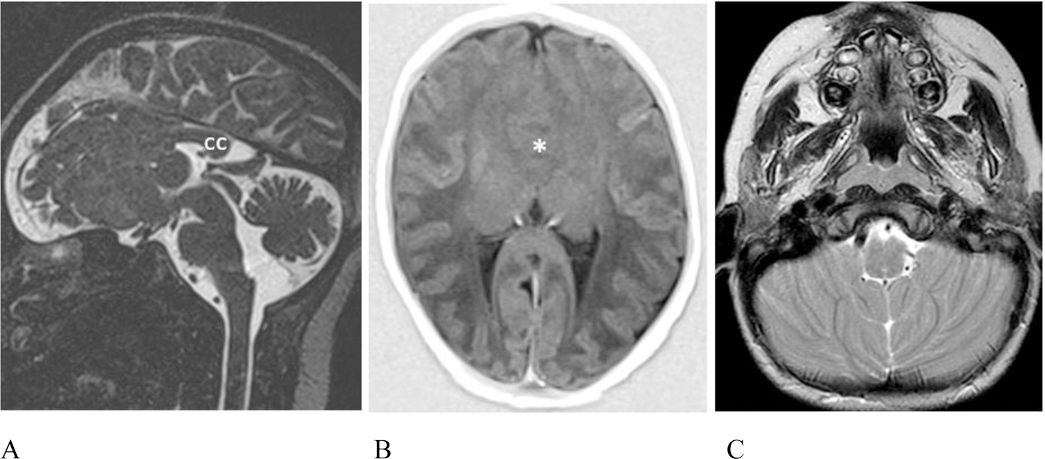
Figure 4.

Additional intracranial anomalies. (A – B) Patient #20, 5 day old female. (A) Sagittal T2 weighted sampling perfection with application-optimized contrasts using different flip angle evolution (SPACE) image reveals absence of the anterior 2/3 of the corpus callosum (cc). The vermis is uplifted and the pons is mildly misshapen and slightly short. (B) Axial T1 inversion recovery MR image shows lobar holoprosencephaly with lack of cleavage of the frontal lobes (asterisk) and dysmorphic lateral ventricles with non-visualization of the frontal horns. The right olfactory bulb was absent (not shown). (C) Patient #7, 4 year old male. Axial T2 weighted MR image shows mildly malformed cerebellar hemispheres which are coapted in the midline and the inferior vermis which is hypoplastic is not seen as expected.
