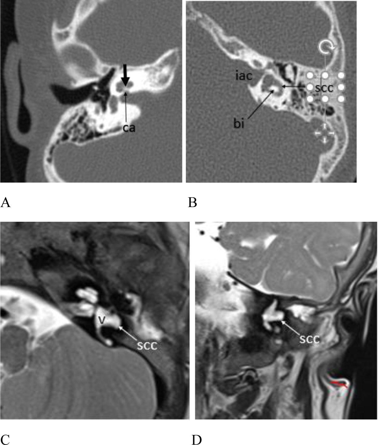
Figure 6.

(A – B) CT and (C – D) MR exams showing inner ear anomalies. (A) Patient #4, 6 year old female, axial CT image right temporal bone. The cochlear modiolus is thickened (short arrow) and there is mild stenosis of the cochlear aperture (arrow ca). (B) Patient #3, 8 year old female, axial CT image left temporal bone. The horizontal SCC lumen is mildly widened anteriorly and laterally (arrow scc), and the bone island (arrow bi) between the SCC and the vestibule is misshapen. Note the widened and somewhat bulbous appearance of the internal auditory canal (iac). Figure 6. cont. Patient #17, 15 month old female. (C) Axial and (D) coronal T2 weighted MR images show malformation of the horizontal SCC (arrow scc) which appears globular anteriorly and deficient posteriorly with absence of the bone island between the vestibule (v) and the horizontal SCC.
