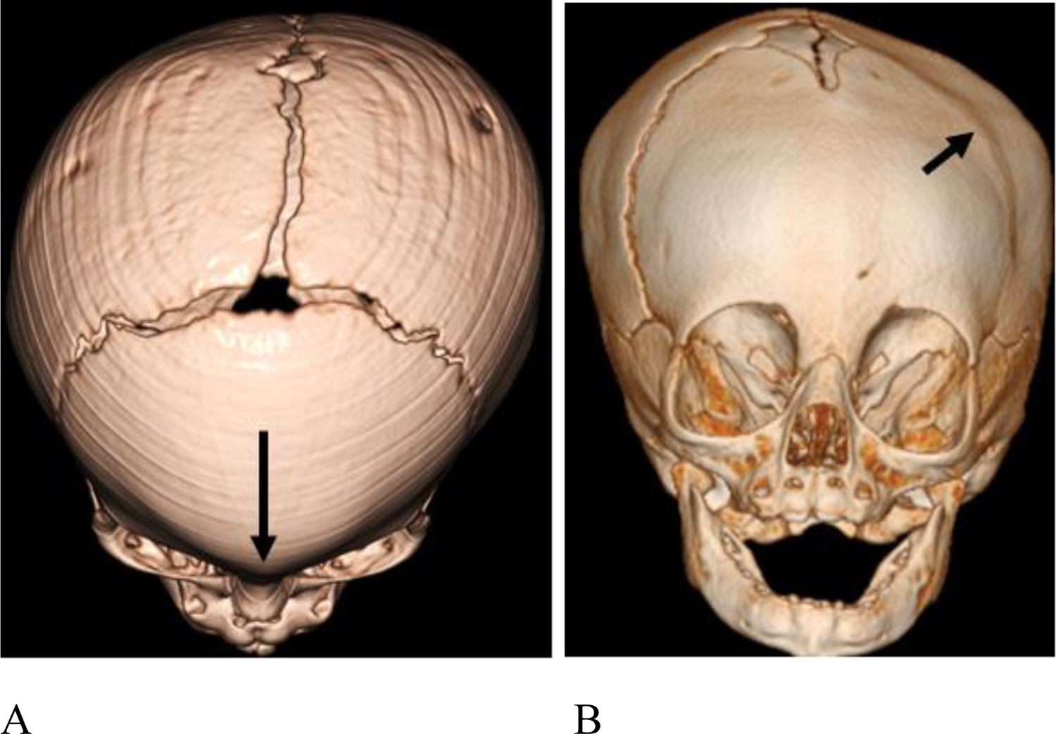
Figure 7.

3D CT images demonstrating craniosynostosis. (A) Patient #3, 7 month old female. There is trigonocephaly with a pointed appearance of the frontal bones in the midline due to premature fusion of the metopic suture (long arrow). There is associated hypotelorism (not shown). (B) Patient #14, 6 month old female with left frontal plagiocephaly due to premature fusion of the left coronal suture (short arrow).
