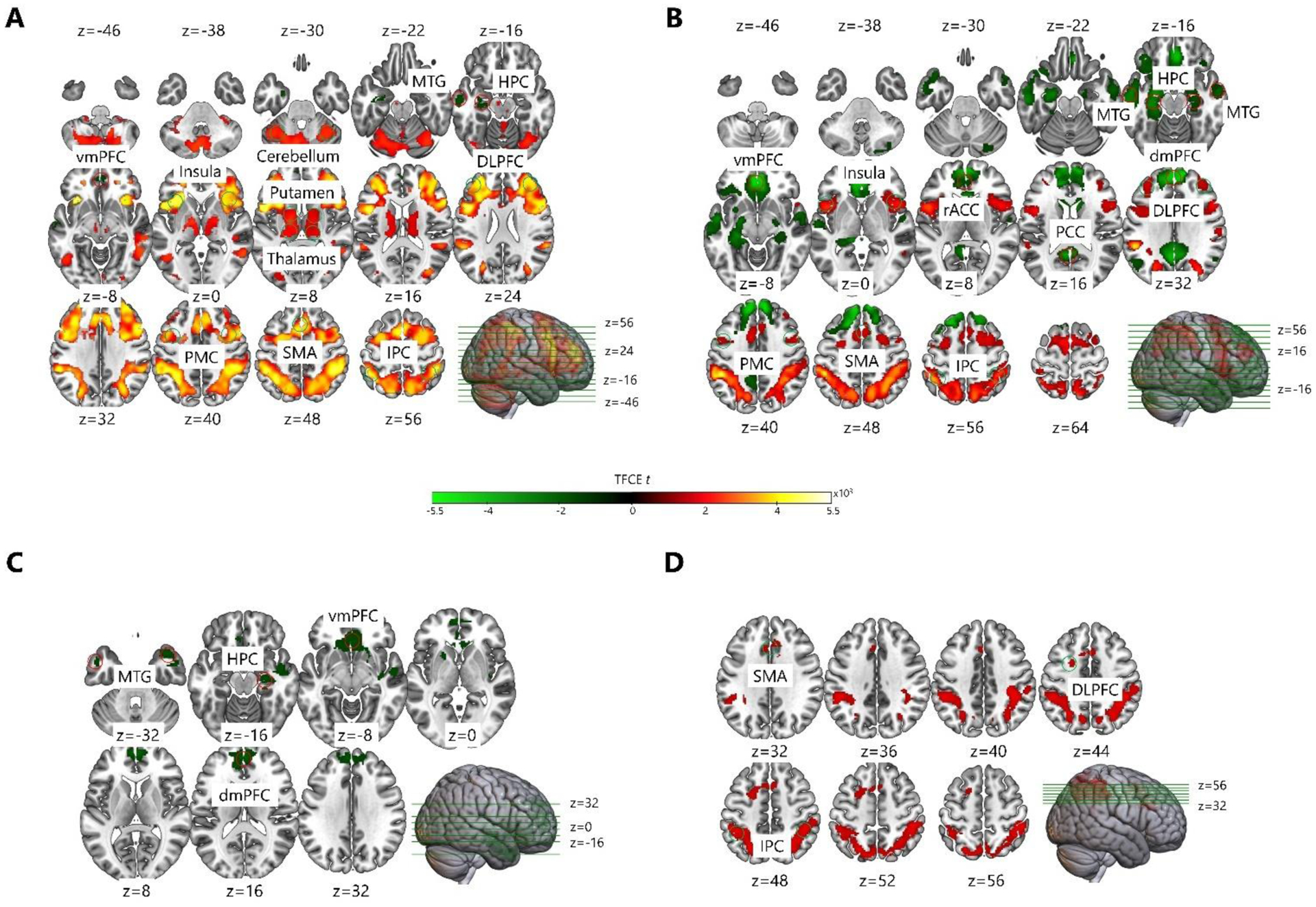
Fig. 5. Brain activation on sequence relative to random trials for the nap condition.

A) During the pre-nap encoding phase, young adults showed increased brain activation in DLPFC, PMC, SMA, IPC, insula, putamen, pallidum, thalamus, and cerebellum, and decreased brain activation in vmPFC, left MTG, and left HPC. B) During the post-nap delayed test phase, young adults showed increased brain activation in DLPFC, PMC, SMA, IPC, and insula, and decreased brain activation in dmPFC, vmPFC, rACC, PCC, MTG, and HPC. C) During the pre-nap encoding phase, older adults showed decreased brain activation in dmPFC, vmPFC, MTG, and right HPC. D) During the post-nap delayed test phase, older adults showed increased brain activation in left DLPFC, SMA, and IPC. DLPFC – dorsolateral prefrontal cortex; dmPFC – dorsomedial prefrontal cortex; vmPFC – ventromedial prefrontal cortex; rACC – rostral anterior cingulate cortex; PCC – posterior cingulate cortex; PMC – premotor cortex; SMA – supplementary motor area; MTG – middle temporal gyrus; IPC – inferior parietal cortex; HPC – hippocampus.
