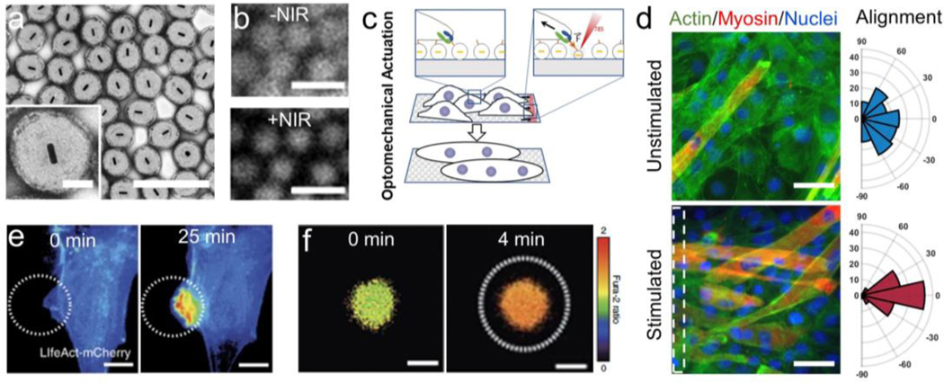
Figure 12: Photoresponsive MAMs direct cell behavior.
a) Optomechanical actuators (OMAs, as described in Section 4.2.1) are gold-pNIPMAm composite nanoparticles that contract when exposed to NIR light. TEM image, scale bar = 1 μm, inset scale bar = 200 nm. b) Fluorescent labeling of OMAs provides evidence particle collapse when exposed to NIR light. Scale bar = 1 μm. c) These MAMs can be modified to facilitate cell attachment, applying force with high spatial precision to study the role of mechanics in cell activity across a variety of cell types. d) Repeated stimulation of myoblasts over 5 days enhanced markers of maturation (left), such as myosin expression (red) and multinucleation (nuclei, blue), as well as cellular alignment (actin, green; histograms, right). Scale bar = 50 μm. e) Fibroblast cells responded to short-term mechanical stimulation by extending in the direction of the stimulus, and increasing actin polymerization. Scale bar = 10 μm. f) Mechanical stimulation of T cells was able to significantly enhance Fura-2 calcium signaling, an important marker of T cell activation. Scale bar = 5 μm. Figures readapted with permission from: a, c-d) Ramey-Ward et al. (2020) ACS Appl. Mat. Inter. Copyright 2020 American Chemical Society [57]; b,e-f) Liu et al. (2016) Nat. Meth. Copyright 2016 Springer-Nature [26].

