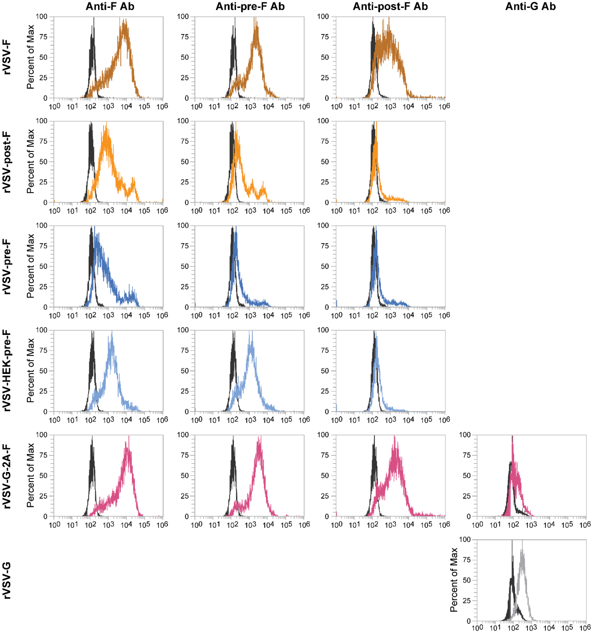
Figure 3. Protein expression on the surface of L929 cells infected with rVSV-F, rVSV-post-F, rVSV-HEK-pre-F, rVSV-G-2A-F, or rVSV-G.

L929 cells were infected for 12 hours with an moi of 5 and protein expression level of F or G protein was assessed via flow cytometry, using an anti-F antibody (Motavizumab), an anti-pre-fusion-F antibody (D25), an anti-post-fusion-F antibody (2F), or an anti-G antibody (m131–2G). Black overlays on the histograms represent unstained, infected L929 cells, while the colored overlays represent stained and infected L929 cells.
