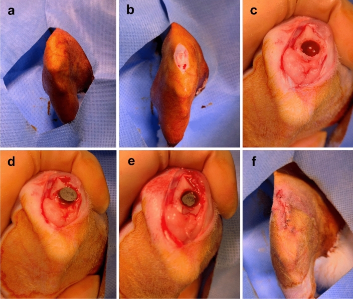
Figure 6.
Surgical procedure for implanting specimens into rabbit trochlear grooves. (a) The right knee of each rabbit was shaved and sterilized with povidone–iodine. (b) In the supine position, the right legs were incised longitudinally from 2 cm above the knee joint to 1.5 cm below. (c) A hole with a 6 mm trephine burr was created on the proximal side of the trochlear groove. (d) A specimen was placed in the hole with the porous surface facing the cancellous bone. (e) The specimen was gently impacted to facilitate contact with the cancellous bone. (f) Patella reduction was performed and the incision was repaired.

