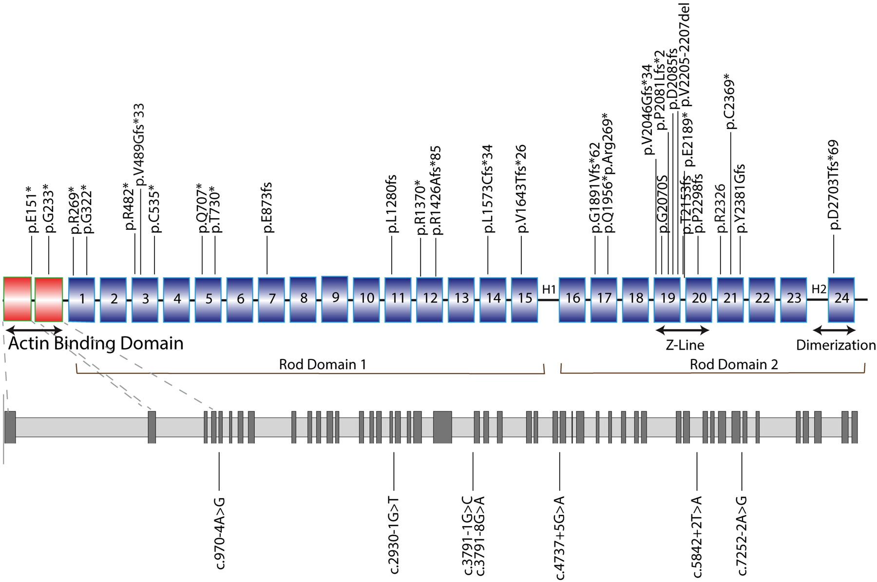
Figure 1. Mapping of FLNCtv variants.

Diagrams representing the structure of FLNC and the distribution of FLNCtv variants. FLNCtv were distributed across all gene domains. However, clusters were noted in the Actin Binding Domain (ABD domains 1 and 2) and in the Z-disk region (Ig-like 19–21) of the Rod Domain 2. Nonsense mutations, and insertion/deletion (indel) variants are indicated in the upper scheme, splice site mutations in the lower (gray) scheme. 1 to 24, Ig-like domains; H1 and H2, hinge domains.
