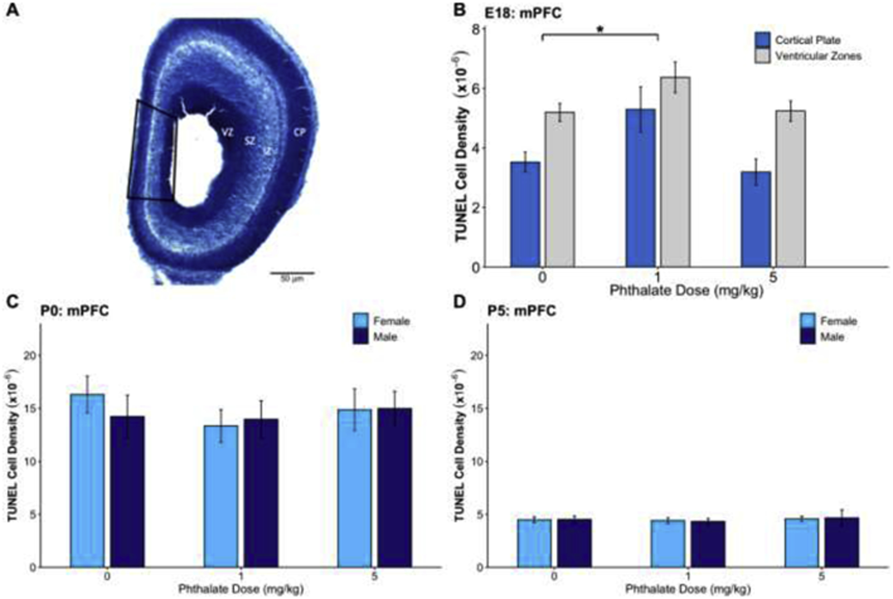
Fig. 3.

The effects of prenatal phthalate exposure on apoptosis. (A) A Nissl-stained section of the E18 cortex depicting the ventricular zone (VZ), subventricular zone (SZ), intermediate zone (IZ), and cortical plate (CP) along with the sampling region of mPFC outlined in black. (B) Prenatal phthalate exposure at the 1mg/kg dose led to increased apoptosis in the mPFC on E18 in the cortical plate and ventricular zones. There was no effect of phthalate exposure on TUNEL density in the mPFC on (E) P0 or (F) P5. *p < 0.05
