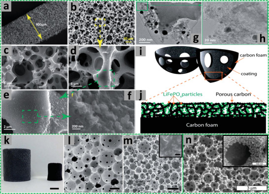
Figure 14.

a,b,d–f) SEM images of LiFePO4 NPs‐coated carbon foam at different magnifications and c) a bare carbon foam. g,h) TEM images confirm the presence of LiFePO4 NPs. i,j) Cartoons show the entrapment of LiFePO4 NPs into the pores of carbon to form a LiFePO4–carbon composite layer. Adapted with permission.[ 237 ] Copyright 2014, Royal Society of Chemistry. k) Reduced graphene oxide (rGO)‐stabilized PDVB polyHIPE monolith (left) and the resulting carbon monolith after carbonization (right), scale bar 5 mm; l) SEM image of PDVB monolith, scale bar 100 µm; m) SEM image of the carbon polyHIPE monolith and the inset image showing rGO platelets, scale bar 100 µm and inset scale bar 400 nm. Adapted under a Creative Commons Attribution 3.0 Unported Licence.[ 241 ] Copyright 2018, The Author(s), published by the Royal Society of Chemistry. n) SEM images showing the HIPE‐templated porous carbon and the mesoporous pore wall (in the inset). Reproduced with permission.[ 233 ] Copyright 2010, American Chemical Society.
