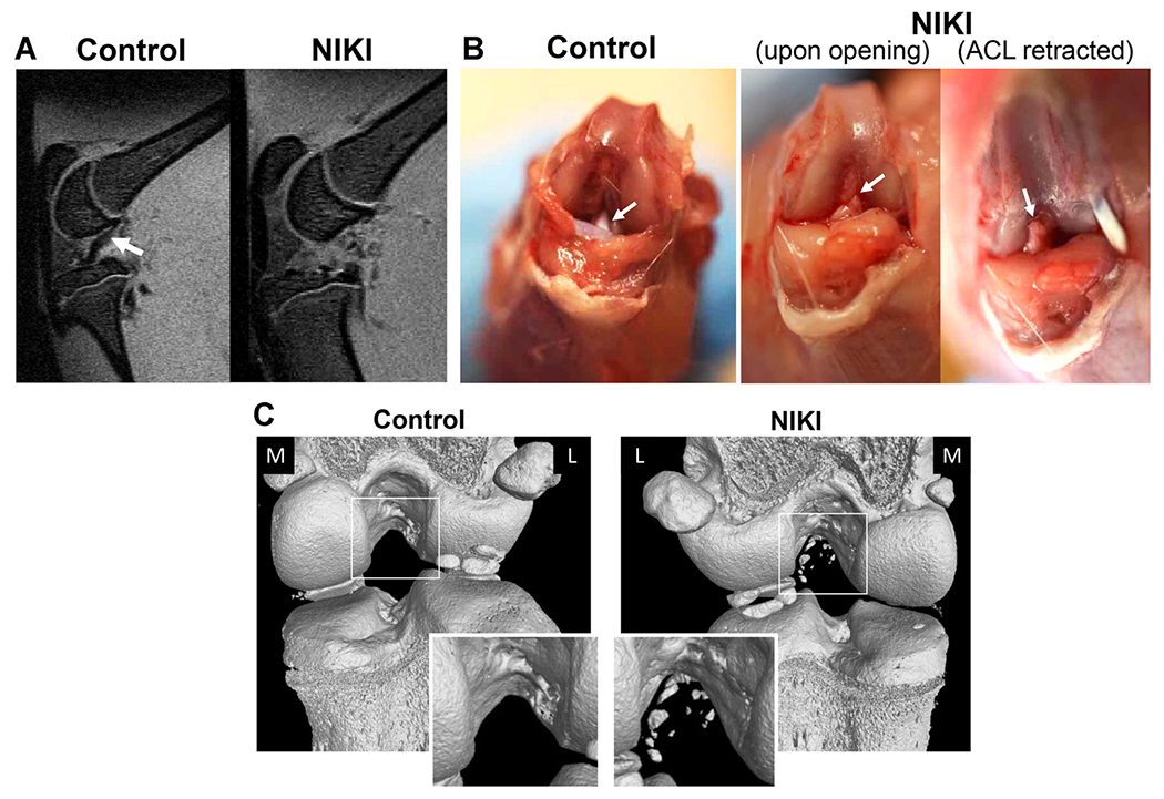
Figure 2.

Confirmation of ACL rupture in animals euthanized immediately post-injury. (A) Sagittal view of the knee by MRI shows the ACL in the contralateral control knee (white arrow), which is not visible in the same view of the injured knee. (B) Appearance of the ACL by gross dissection, whereby in injured knees, the ACL (white arrow) had been torn from the femoral attachment site and was fully retractable. (C) Bone condition and tear type identification via nanoCT immediately post-injury. The nanoCT-imaged knees are shown from the posterior view. The contralateral controls are animal-matched in all images. M = medial, L = lateral. [Color figure can be viewed at wileyonlinelibrary.com]
