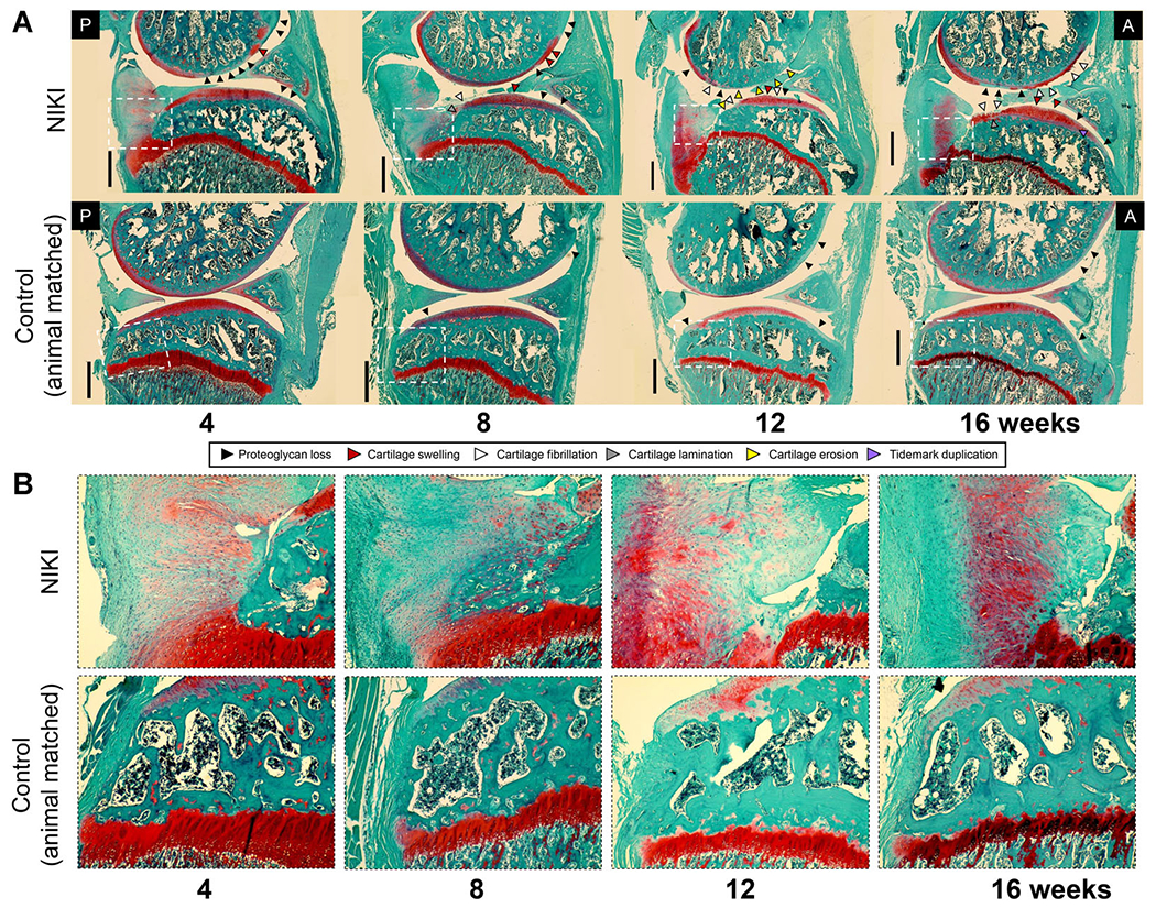
Figure 4.

(A) Histological assessment of the whole joint stained with Safranin-O/fast green. Images were taken in the medial compartment at ×4, then stitched together to view a larger region of the joint. (B) ×10 zoom on the posterior tibial plateau (region identified by white dotted lines in (A)). All images were taken at the same microscope settings. A = anterior, P = posterior. Scale bar = 1 mm. [Color figure can be viewed at wileyonlinelibrary.com]
