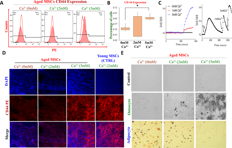
Fig. 2.
Role of calcium in differentiation and surface marker expression on young and old MSCs. A Immunophenotyping assay was done to study the CD44 surface marker expression after various calcium treatment (0 mm Ca2+, 2 and 5 mM Ca2+ for 3 days). B, shows the histogram showing the expression of the CD44 surface marker expression in varying dose of Ca2+ treatment. C Representative traces showing changes in intracellular Ca2+ levels after various Ca2+ treatment. D represent CD44 expression in aged MSCs in various Ca2+ concentrations. Young MSCs grow in in 2 mM Ca2+ was used as control. E Adipocyte and osteocyte differentiation assay was performed to study the stem cell differentiation potential of aged MSCs after treatment with different doses of Ca2+.

