Fig. 3.
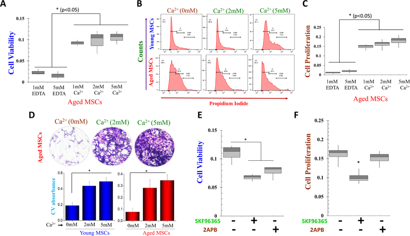
Role of calcium on aged MSCs cell viability and cell proliferation after increasing calcium concentrations. A MTT assay was performed to investigate calcium’s effect on aged MSCs cell viability. Aged MSCs cells were treated with 5 mM EDTA (mimic 0 mM Ca2+) and increasing Ca2+ concentrations for 1 day. B Cell cycle assay was done to study the phases of the cell cycle after Ca2+ treatment. The bar graph shows the increment of the cell growth phase, i.e., S phase, and diving phase, i.e., G2/M phase with increasing Ca2+ conc. C BrdU assay was performed to study the cell proliferation of MSCs after treatment with 5 mM EDTA and different doses of Ca2+ for and 3 days. D The colony-forming unit (CFU) assay shows the stem cell potential after the treatment of exogenous Ca2+. CFU image and graph show increased stem cell potential with increasing Ca2+ concentration. E MTT assay and F BrdU assay were performed to investigate calcium’s proliferative effect. MSCs cells were treated with SKF 96,365 and 2-APB for 24 h. Bar graphs depicts average ± SE in 3–4 separate experiments, NS = non-significant, *p ≤ 0.001 (Student’s t test).

