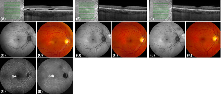
Fig. 1.

Multimodal imaging of a 47‐year‐old man with chronic central serous chorioretinopathy who was treated twice with half‐dose photodynamic therapy (PDT) in the PLACE trial. At baseline visit of the PLACE trial (before PDT; A–E), subretinal fluid and discontinuity of the external limiting membrane (ELM) and ellipsoid zone (EZ) were visible on optical coherence tomography (OCT; A). In addition, a focal leakage point was visible on fluorescein angiography (FA; D) and hyperfluorescent abnormalities were present on indocyanine green angiography (ICGA; E). Both at baseline visit of the current study (8 months after treatment within the PLACE trial; F–H) and at final visit of the current study (20 months after treatment; I–K), subretinal fluid had resolved and the ELM and EZ were no longer discontinuous on OCT (F, I). Fundus autofluorescence and fundus photography did not markedly change between baseline visit and final visit of this study (G, J and H, K, respectively). FA and ICGA were not obtained at baseline visit and final visit of this study due to the absence of subretinal fluid.
