FIGURE 4.

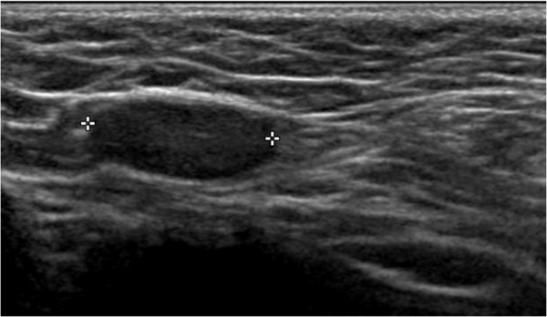
Ultrasound images of an inguinal cryptorchid testes using high‐frequency (12 MHz) linear probe. The testicle appears hypoechoic

Ultrasound images of an inguinal cryptorchid testes using high‐frequency (12 MHz) linear probe. The testicle appears hypoechoic