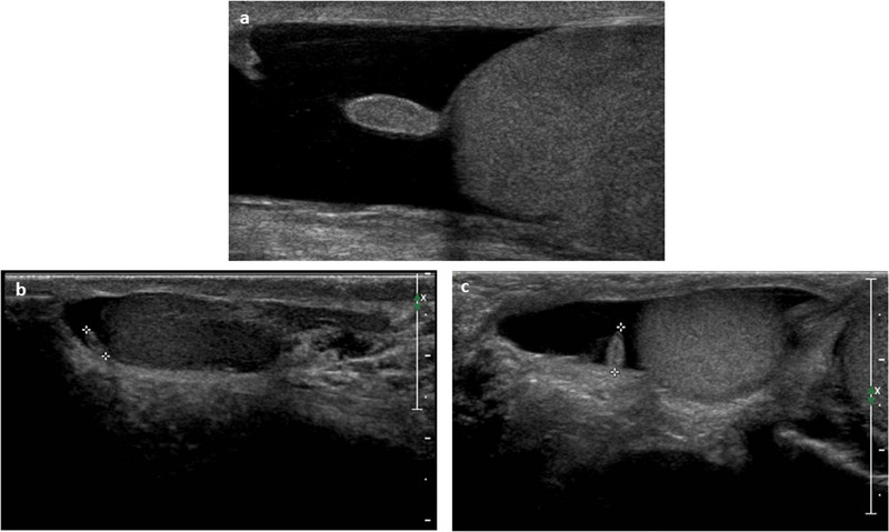
FIGURE 6.

Ultrasound images of (A, B) testicular and (C, D) epididymal appendages using high‐frequency (12 MHz) linear probe. They appear oval in shape and isohypoechoic; visualization may be aided by the presence of a reactive hydrocele

Ultrasound images of (A, B) testicular and (C, D) epididymal appendages using high‐frequency (12 MHz) linear probe. They appear oval in shape and isohypoechoic; visualization may be aided by the presence of a reactive hydrocele