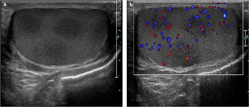
FIGURE 8.

Ultrasound images of testicular recurrence of leukemia using high‐frequency (12 MHz) linear probe. (A) Several hypoechogenic foci of leukemic infiltration (recurrence of acute lymphatic leukemia). (B) The color Doppler US results, which shows an increased blood flow in each lesion
