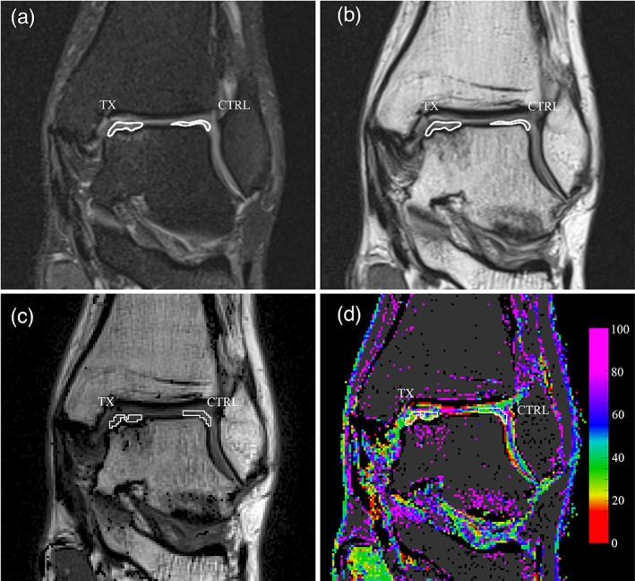
FIGURE 2.

Definition and transposition of the volumes of interest (VOIs). (a) Manual delineation of the surgically treated area (TX) and cartilage control area (CTRL), on the coronal PD‐TSE sequence performed in Horos software. The VOIs are shown as contour lines on the interpolated rendering of the image. (b) The same VOIs in the first echo signal of the multi‐echo sequence, after optional alignment correction. (c) The VOIs in the first echo signal imported in MATLAB software. VOIs are delineated in white. (d) The same VOIs transposed on the T2 map, also shown in white. The image was produced without interpolating color between voxels.
