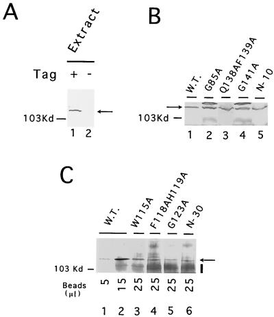
FIG. 5.
Analysis of mutant protein expression. (A) Whole-cell extracts were prepared from strains whose Est2p was either tagged (+) or untagged (−) with the IgG binding domain of protein A and subjected to Western analysis. For each extract, 300 μg of total protein was examined. The position of the protein A-tagged Est2p is indicated by an arrow. (B) Est2p levels in whole-cell extracts from wild-type (W.T.) or mutant strains were analyzed by Western blotting. For each extract, 500 μg of total protein was examined. The position of the protein A-tagged Est2p is indicated by an arrow. (C) Est2p levels in whole-cell extracts from wild-type (W.T.) or mutant strains were assessed by affinity precipitation followed by Western analysis. For each extract, 4 mg of total proteins was incubated with 40 μl of IgG-Sepharose resin with gentle agitation at 4°C for 16 h. The beads were washed extensively, and the indicated amount of Sepharose was boiled in SDS-PAGE loading buffer. The eluted proteins were then subjected to immunoblotting. The position of the protein A-tagged Est2p is indicated by an arrow. The increased background (marked by a vertical bar) was due to IgG that eluted from the beads and that cross-reacted with the secondary antibody.

