
FIG. 3.
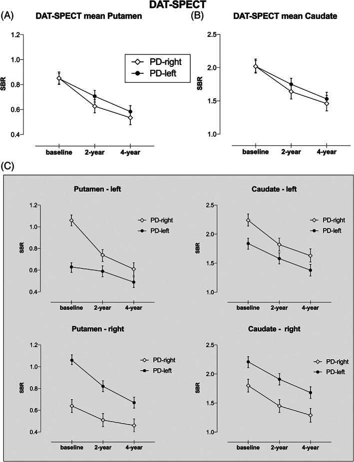
DAT‐SPECT imaging progression in PD with predominant dopaminergic putaminal deficit on the left (PD‐left) versus the right side (PD‐right), namely in the (A) mean putamen, (B) mean caudate, and (C) left/right side of the putamen and caudate nuclei. DAT, dopamine transporter; PD, Parkinson's disease; SPECT, single‐photon emission computed tomography; SBR, specific binding ratio.