
FIGURE 9.
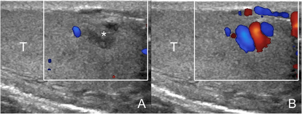
Intratesticular varicocoele. Images obtained at rest (A) and during Valsalva's manoeuver (B). At rest (A) a hypoechoic lesion is seen (asterisk) resembling a tumor. During Valsalva (B) enlarged intratesticular veins with reflux are revealed (T = testis)