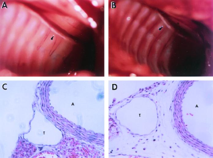
FIG. 3.
Anatomy and histology of thoracic duct in α9−/− mouse and a wild-type littermate at 8 days of age. Presented are photographs of the opened thorax of α9+/+ (A) and α9−/− (B) mice, showing the presence of a visible thoracic duct in both groups (the white tubular structure along the spine denoted by arrowheads). (C and D) H&E-stained sections of the region including the thoracic duct (T) and the adjacent aorta (A) in α9+/+ (C) and α9−/− mice (D), demonstrating edema and extravascular lymphocytes surrounding the thoracic duct in α9−/− but not in α9+/+ mice.

