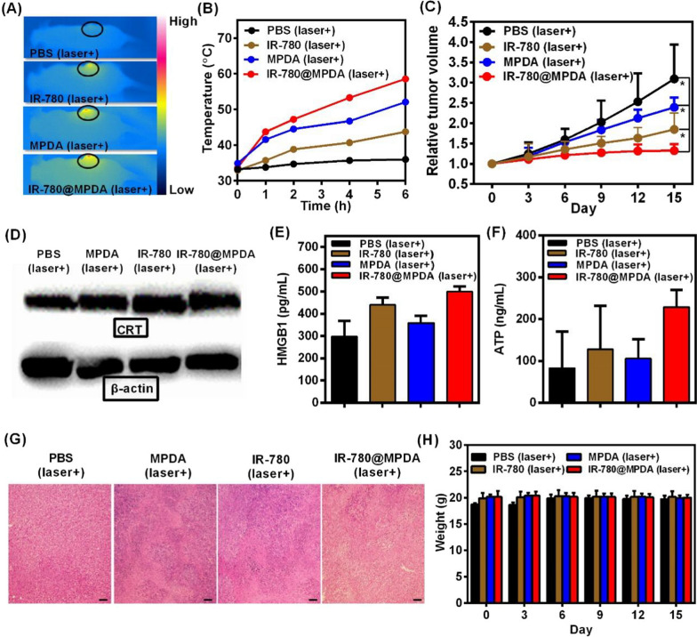
Fig. 5.
A The representative thermal images of tumor-bearing mice after i.v. injection of PBS, IR-780, MPDA, and IR-780@MPDA at 6 h. B Tumor temperature elevation under laser irradiation (808 nm, 1 W/cm2, and 300 s) in mice after i.v. injection of PBS, IR-780, MPDA, and IR-780@MPDA at 1, 2, 4, and 6 h. C Relative tumor volume of tumor-bearing mice after indicated treatments. The tumor volumes were normalized to their initial sizes. Asterisk * indicated p < 0.05. D Western blot analysis of CRT expression of tumor after different treatments. E Detection of HMGB1 release in vivo. F Detection of ATP secretion in vivo. G H&E staining of tumors after indicated treatments, scale bars: 100 μm. H Changes in the body weight of mice after indicated treatments

