
Figure 4.
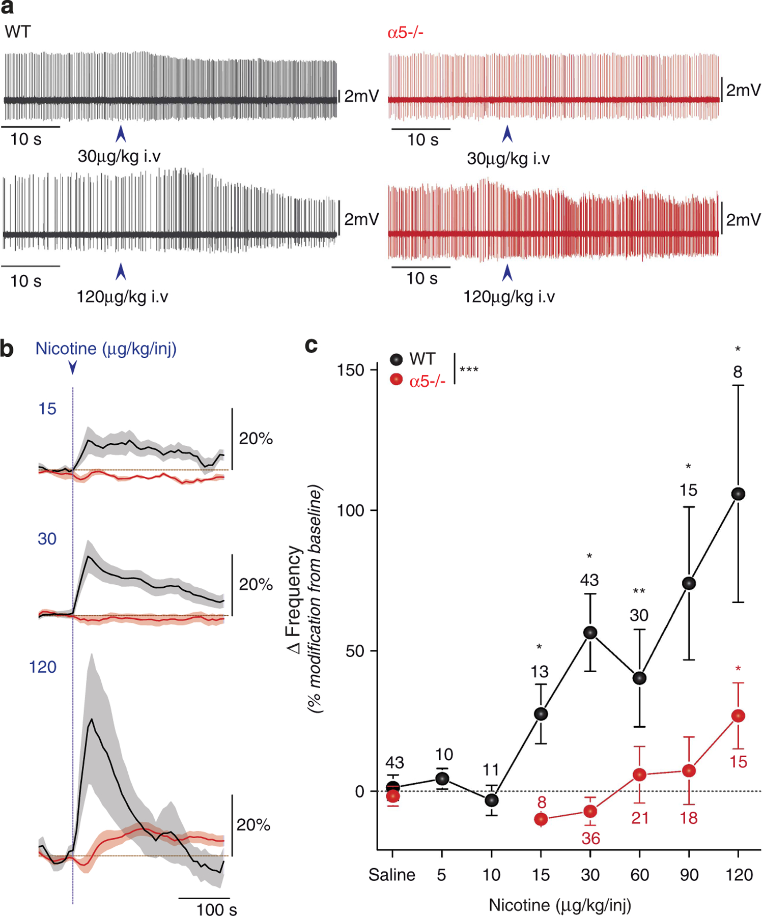
Nicotine-elicited increase in ventral tegmental ares (VTA) DA cell firing is shifted to higher doses in α5−/− mice. (a) Typical electrophysiological recording depicting the changes in firing pattern elicited by 30 μg kg−1 or 120 μg kg−1 intravenous (i.v.) nicotine injection (arrow) in wild-type (WT) and α5−/− mice. (b) Mean ± s.e.m DA cell firing frequency increase after injection of the indicated nicotine concentration, in WT and α5−/− mice. (c) Rightward shift in the dose-response curve of nicotine-elicited DA cell activation in α5−/− mice. Mean ± s.e.m of increased variation from baseline in firing frequency for WT and α5−/− mice injected with the indicated nicotine concentrations. WT: black; α5−/−: red. ***P<0.001, Kruskall–Wallis test; *P<0.05, **P<0.01 Wilcoxon test. n, number of recorded neurons.