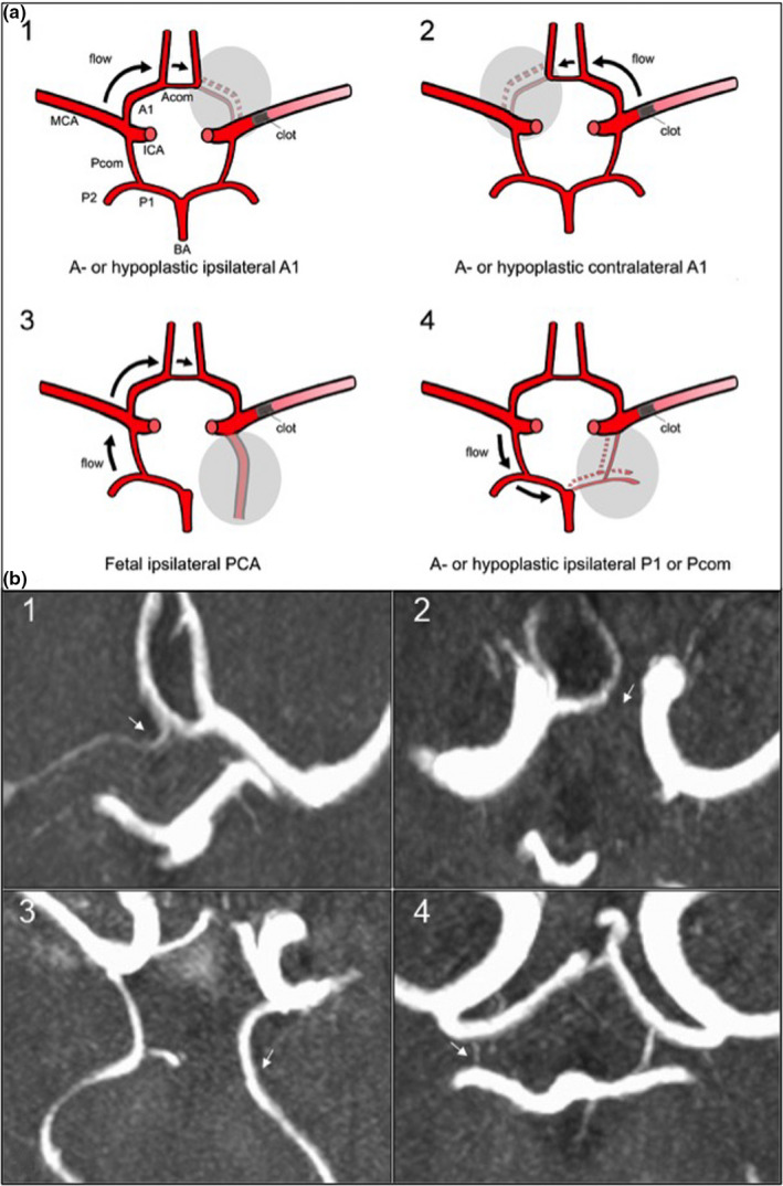
FIGURE 1.

Scheme of vascular models of circle of Willis (CoW) variants (a) and visualization in time‐of‐flight magnetic resonance angiography (TOF‐MRA). (b) Axial magnetic resonance images of acute ischemic stroke patients performed on admission showing the CoW in 3D‐multi‐slab TOF‐angiography in the maximum intensity projection (MIP). CoW variant indicated with a white arrow on each image. 1: Hypoplastic A1‐segment ipsilateral to M1‐occlusion on the right side; 2: aplastic A1‐segment contralateral to M1‐occlusion on the right side; 3: full fetal PCA ipsilateral to M1‐occlusion on the left side; 4: hypoplastic Pcom ipsilateral to M1‐occlusion on the right side. Abbreviations: Acom, anterior communicating artery; ICA, internal carotid artery; MCA, middle cerebral artery; PCA, posterior cerebral artery; Pcom, posterior communicating artery
