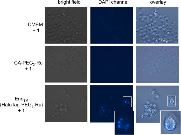
Figure 6.
Fluorescence microscopy and bright‐field images of J774A.1 monocytes treated with StrepEncSM{HaloTag‐PEG3‐Ru} + 1, CA‐PEG3‐Ru (5) + 1 and 1 only (negative controls). Cells were seeded and treated with 1 μM StrepEncSM{HaloTag‐PEG3‐Ru} in DMEM overnight. Subsequently, 1 (20 μM) was applied to the cell supernatant. Following incubation and washing, images were acquired with a digital inverted microscope. Fluorescence microscopy images of monocytes treated with encapsulin show fluorescence from uncaging product 2 that is localized within distinct vesicles. Fluorescence microscopy images show no blue fluorescence in cells treated with 1 only. Key: Dulbecco's Modified Eagle Medium (DMEM).

