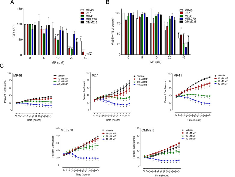
Fig. 1 .
MF inhibits functionality, growth capacity, and viability of UM cell lines in a concentration-related manner. Graphs represent the level of cellular functionality or viability, respectively as detected via a CCK8 colorimetric assay (A) or Trypan Blue exclusion assay (B) after cells were treated with increasing concentrations of MF (0, 5, 10, 20, or 40 μM) for 72 h. C Growth curves obtained through Incucyte live cell imaging system, tracking cellular confluency. In A and B, data were analyzed using two-way ANOVA followed by Dunnett’s multiple comparison test. In C, data were analyzed using repeated measures ANOVA followed by Tuckey’s multiple comparison test.* Indicates p < 0.05, ** indicates p < 0.01, whereas *** indicates p < 0.001 compared against vehicle-treated controls

