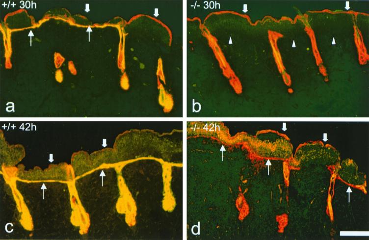
FIG. 3.
Immunofluorescence analysis of reepithelialization after tape stripping. All samples were stained with antibodies to MK14 (red) and MK6 (yellow). The dead old basal layer is still present on top of the crust (thick arrows) and is positive for MK14 but negative for MK6. (a) MK6a+/+ animals show some areas of reepithelialization 30 h after tape stripping. A single-cell layer, positive for MK6, is apparent under the crust (thin arrows). Note the induction of MK6a in the ORS of the hair follicles. (b) MK6a−/− mice show no reepithelialization from the hair follicles 30 h after tape stripping. Arrowheads, boundary of crust and dermis; thick arrows, old basal layer on top of the crust. (c) MK6a+/+ samples taken 42 h after tape stripping show a continuous epithelium of two to four cell layers under the crust (thin arrows). (d) MK6a−/− samples taken 42 h after tape stripping show a discontinuous mostly single-cell layer epithelium under the crust (thin arrows). Note that the follicular cells that reach the dermal surface do not induce MK6b until a suprabasal layer is present. Scale bar, 100 μm.

