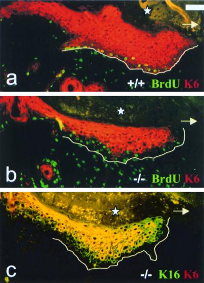
FIG. 5.
Immunofluorescence analysis of full-thickness wounds. Arrow, direction of migration of the epithelial tongue; star, crust covering the wound; line, boundary of the migrating epithelial tongue and the granulation tissue. (a) MK6a+/+ 5-day-old full-thickness wound. All layers of the migrating epithelial tongue are MK6 positive (red). Note that in order to show a well-preserved wound, fixed tissue, on which this MK6 antibody results in weaker staining of the basal layers, is shown; however, this is not the case on unfixed frozen sections (not shown). Proliferating keratinocytes in the bottom layers of the epithelial tongue are BrdU (FITC) and MK6a (TxRed) positive and therefore appear to have yellow nuclei. The proliferating cells in the granulation tissue are only BrdU positive, and the nuclei appear green. (b) MK6a−/− 5-day-old full-thickness wound. Note that the BrdU-positive, proliferating keratinocytes are negative for MK6b. (c) Double staining for MK16 (FITC) and MK6 (TxRed) in MK6a−/− animals reveals that MK16 is induced in cells below those expressing MK6b. Scale bar, 50 μm.

