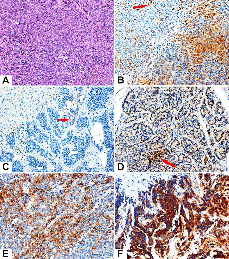
Figure 1.
Microscopic features of the NENs examined in the study. (A) LCNEC. (B) High-grade urothelial carcinoma served as positive control. Cytoplasmic and membranous positive expression was observed within tumor cells, as well as positive endothelial cells (red arrow). (C) Liver metastasis from a well-differentiated G2 NET with IRS=0 (the nuclear staining of rare tumor cells is not counted). (D) Well-differentiated G1 NET, IRS=8. A small group of positive intra-tumoral lymphocytes is marked with red arrow. (E) LCNEC, IRS=9. (F) SCNEC with intensely positive cytoplasm of the tumor cells, IRS=12. HE staining: (A) ×100. Anti-CXCR4 chemokine receptor antibody immunomarking: (B–F) ×400. CXCR4: C-X-C motif chemokine receptor 4; HE: Hematoxylin–Eosin; IRS: Immunoreactivity score; LCNEC: Large-cell neuroendocrine carcinoma; NEC: Neuroendocrine carcinoma; NENs: Neuroendocrine neoplasms; NET: Neuroendocrine tumor; SCNEC: Small-cell neuroendocrine carcinoma

