
Fig. 5.
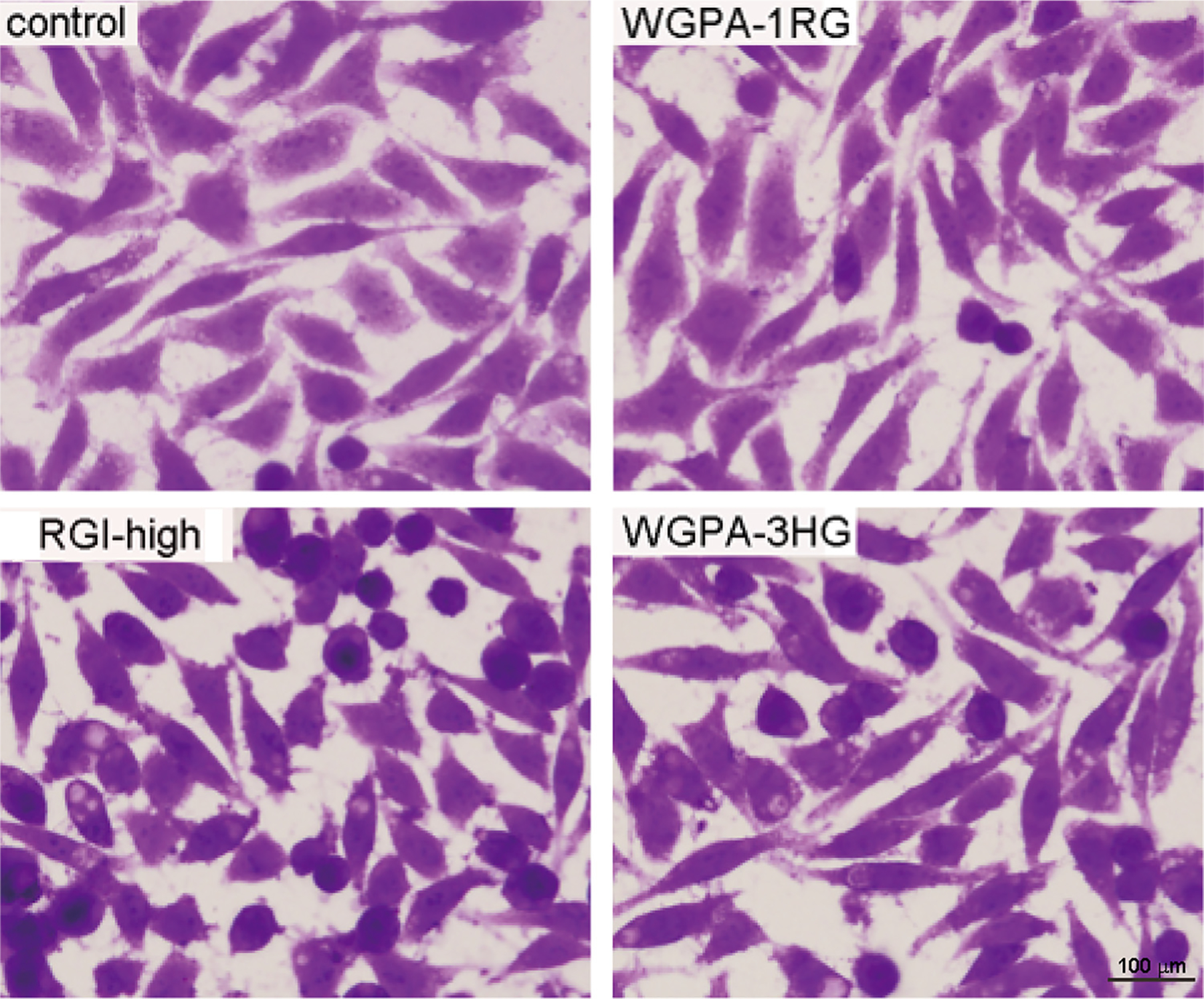
Pretreatment of cells by HG- and RGI-rich pectins altered cell morphology. L-929 cells treated without (control) or with 0.5 mg/ml WGPA-1RG, RGI-high, WGPA-3HG for 24 h were fixed, stained with crystal violet and visualized under microscope at 200× magnifications. This experiment was repeated twice. The representative images from total 32 fields under each condition were shown.