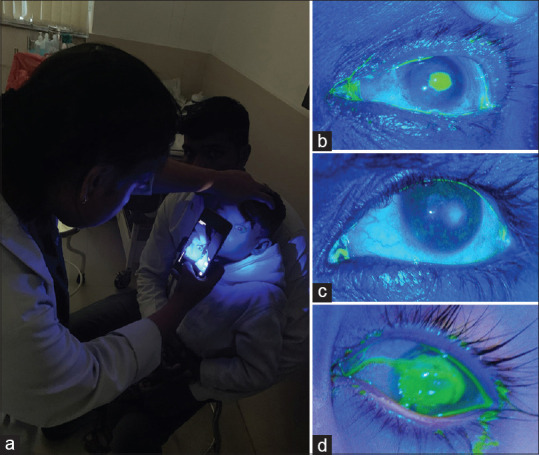
Figure 3.

(a) Fluorescein-stained cornea of a child being photographed using a cobalt blue light unit (C-BLU) filter mounted on a smartphone. (b) Still images captured using C-BLU technique during remote eye camp that demonstrate stain-positive central corneal ulcer. (c) Diffuse corneal staining due to chronic dry eye. (d) A 1-year-old child presented with xerophthalmia which was documented using C-BLU
