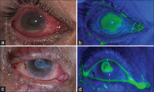
Figure 4.

(a) Large central corneal ulcer with hypopyon seen under diffuse illumination. (b) The same ulcer as in (a) photographed using the C-BLU filter system after staining with fluorescein. (c) A deep central corneal ulcer with descemetocele under diffuse illumination. (d) C-BLU system used to capture fluorescein-stained ulcer seen in (c)—the characteristic “green donut” appearance of descemetocele with a ring of stain-positive stroma surrounding a dark center can be seen
