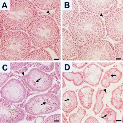
FIG. 1.
Histopathology of Egr-deficient testes. (A) Adult wild-type testes contain seminiferous tubules filled with maturing germ cells. The steroid-producing Leydig cells (arrowhead) are located in the interstitial spaces between the tubules. (B) In testes from Egr1-deficient mice, spermatogenesis proceeds normally. However, Leydig cells (arrowhead) are notably atrophic. (C) The testes from adult Egr4-deficient mice show marked disruption of spermatogenesis, high levels of germ cell apoptosis (arrow), and marked Leydig cell hyperplasia (arrowhead). (D) The testes from adult Egr4-Egr1-deficient mice contain small atrophic seminiferous tubules filled with very few maturing germ cells. The majority of cells within the seminiferous tubules consist of aggregated Sertoli cells (arrows). Leydig cells are markedly atrophic (arrowhead). (Bar = 50 μm.)

