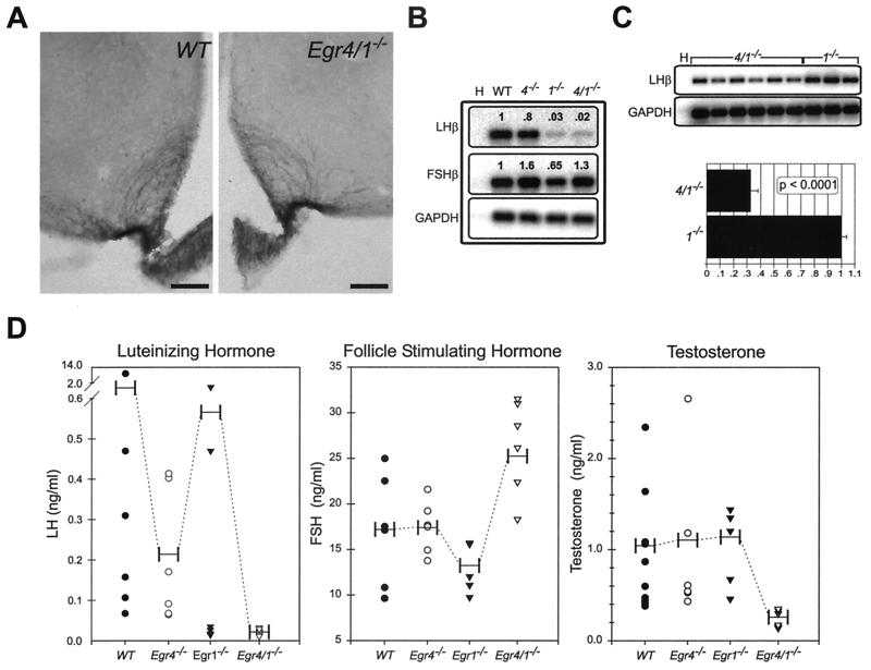
FIG. 4.
Low serum LH levels in Egr4-Egr1-deficient male mice are associated with low serum testosterone levels. (A) Immunohistochemical studies show no alterations in the integrity of GnRH-positive neurons in the hypothalamus (not shown) or their terminals that innervate the median eminence. (B) In the pituitary gland, semiquantitative RT-PCR analysis from pooled samples (n = 3, each genotype) demonstrates extremely low levels of LHβ expression in both Egr1- and Egr4-Egr1-deficient mice that are below the dynamic range of the assay. The levels of FSHβ expression are essentially unaffected (H) (no template water control). (C) Semiquantitative RT-PCR analysis of adult male pituitaries (Egr4-Egr1 deficient, n = 6; Egr1 deficient, n = 3) at increased PCR cycle numbers, but still within the logarithmic range of amplification, demonstrate that LHβ is threefold higher in Egr1-deficient relative to Egr4-Egr1-deficient mice (H) (no template water control). (D) LH serum levels are also different between adult Egr1- and Egr4-Egr1-deficient male mice. In Egr1-deficient mice, there appears to be some pulsatile release of LH, but in Egr4-Egr1-deficient mice, only low steady-state levels are observed. FSH levels are mildly elevated in Egr4-Egr1-deficient adult male mice. Low steady-state levels of LH dramatically affect serum testosterone levels in Egr4-Egr1-deficient mice. Apparently, in male Egr1-deficient mice, there is adequate pulsatile release of LH to maintain testosterone synthesis by Leydig cells (horizontal bars represent mean values).

