
Fig. 1.
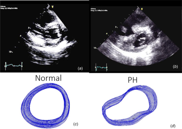
Parasternal short-axis views of the LV in normal (a) subjects and subjects with PH (b) illustrating gross shape deformation in severe PH. Images (c) and (d) show tracings of the endocardial border in all frames acquired over one cardiac cycle. Obvious shape differences are readily perceptible.