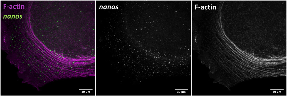
Figure 4.
Micrograph of F-actin arcs and germ plasm RNPs (nanos) in the blastodisc periphery of a fixed 2-cell (45 minutes post-fertilization) zebrafish embryo. As described in Method 3.4, fluorophore-conjugated phalloidin was incorporated with the fixation step to label F-actin (pseudocolored magenta here) followed by in situ hybridization to visualize fluorescein-labeled probes bound to nanos RNA. Image is a 2D projection of a Z-stack acquired on a Zeiss LSM780 confocal microscope.

