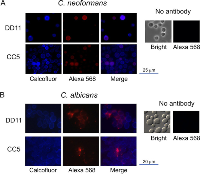
FIG 3.

Reactivity of chitooligomer MAbs with the cell surface of C. neoformans (A) and C. albicans (B). Cell wall chitin was stained with calcofluor white (blue fluorescence), and chitooligomers were stained with an Alexa Fluor 568 secondary antibody (red fluorescence) after incubation with MAb DD11 or CC5. Merge panels illustrate the surface localization of the chitooligomers in more detail. Control systems (no antibody) were not incubated with the primary antibodies. In these systems, fungal cells were observed in bright-field and red fluorescence (Alexa 568) modes.
