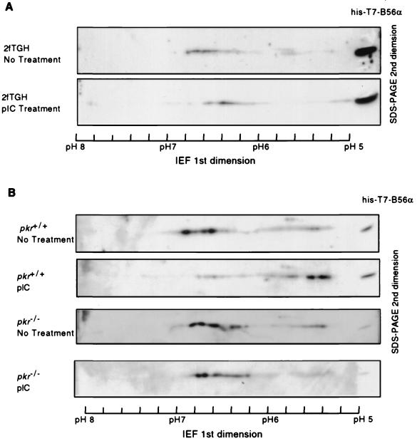
FIG. 4.
Mobility shift of B56α in IEF in response to pIC treatment of the cell. (A) Human 2fTGH cells were transfected with FLAG-B56α expression plasmid DNA pZeoSV-FLAG-B56α. The cells were either treated (bottom panel) or not treated (top panel) with 100 μg of pIC per ml to activate PKR. B56α was immunoprecipitated from 2 mg of cell lysate and separated in the first dimension with IEF in a ready-strip IPG strip (pH range, 5 to 8; Bio-Rad) and in the second dimension with an SDS-polyacrylamide gel. Western blotting with a polyclonal antibody against human B56α was performed to detect B56α. Recombinant His-T7-B56α was loaded at the rightmost side and used as reference for alignment. B56α from 2fTGH cells showed pI values varying from 6.2 to 6.8, but it is very likely that more acidic forms of B56α failed to be detected because of a lower expression level of B56α in 2fTGH cells compared with murine fibroblasts in panel B. (B) pI of FLAG-B56α from pkr+/+ or pkr−/− cells treated or not treated with pIC. The experimental procedure is same as for 2fTGH cells in panel A. The pI values range from 5.3 to 6.8.

