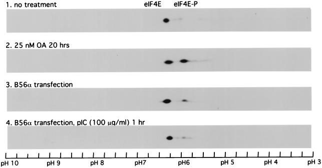
FIG. 9.
IEF–SDS-PAGE analysis of eIF4E phosphorylation in 2fTGH cells. 2fTGH cells were either left untreated (panel 1), treated with 25 nM okadaic acid (OA) for 20 h (panel 2), transfected with B56α expression plasmid DNA pZeoSV-B56α (panel 3), or transfected with pZeoSV-B56α and then treated with 100 μg of pIC per ml for 1 h (panel 4). Cells were lysed, and proteins from 300 μg of cell extract were separated with IEF–SDS-PAGE two-dimensional electrophoresis. eIF4E protein was detected by Western blotting using a monoclonal antibody against rabbit eIF-4E. Phosphorylated eIF4E has a lower isoelectric point and migrates to a lower pH position than unphosphorylated eIF4E.

