. 2021 Oct 25;12(44):14871–14882. doi: 10.1039/d1sc03056f

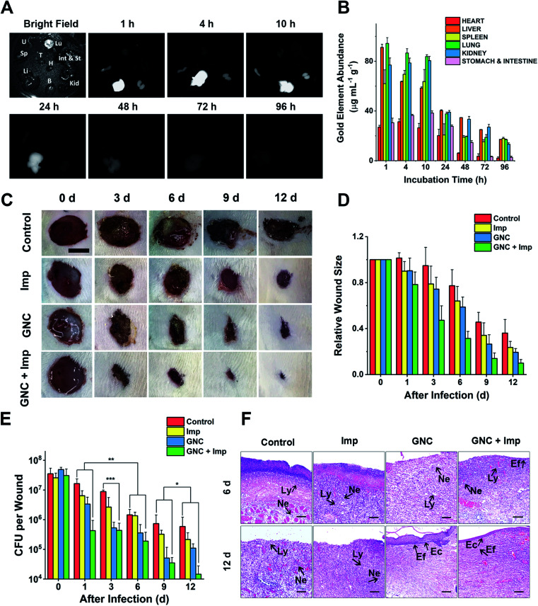
Fig. 6. In vivo experiments demonstrating the clearance time and treatment effect. (A) NIR fluorescence of crucial organs of mice harvested at different time points under bright-field and 808 nm laser radiation (B: brain, H: heart, Int & St: intestine and stomach, Kid: kidneys, Li: liver, Lu: lungs, Sp: spleen, T: thymus and U: uterus). (B) Gold content (average weight) in different organs, harvested at different times post-injection (n = 4). (C) Representative images of changes in wound size in different groups within 12 days post-treatment (scale bar: 10 mm). (D) Comparison of the relative wound size vs. time in different groups: PBS (control), 24 μg mL−1 Imp solution, 64 μg mL−1 GNC solution, and the combination of 24 μg mL−1 Imp (∼0.12 mg kg−1, final concentration) and 64 μg mL−1 GNC solution (∼0.32 mg kg−1, final concentration, same below) (n = 3). (E) Plate coating results of the PBS control, single application, and combined therapy groups (n = 3, *P > 0.05, ** 0.01 < P < 0.05 and ***P < 0.01). (F) The hematoxylin–eosin staining graphs in different groups. After 12 days, the epidermis was fully reconstructed in the GNC + Imp group (Ly: lymphocyte; Ne: neutrophil; Ec: epithelial cell and Ef: elongated fiber).

Fig. 6