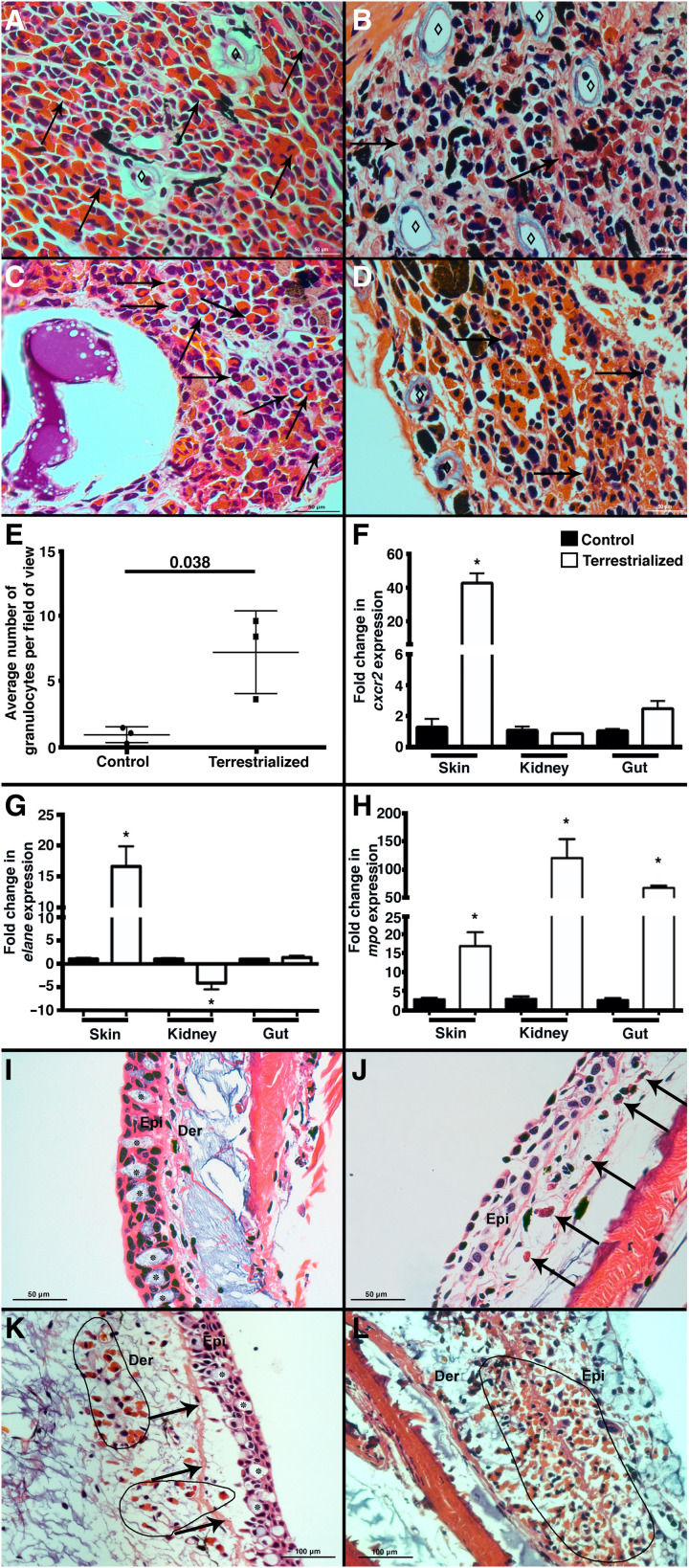
Fig. 1. Lungfish terrestrialization results in the mobilization of granulocytes from reservoir tissues into the integument.
H&E staining of (A) control gut, (B) terrestrialized gut, (C) control kidney, and (D) terrestrialized kidney tissues of African lungfish (n = 3 per group). Black arrows denote examples of granulocytes, and ⋄ denotes lymphatic micropumps. Note the increased abundance of brown and black pigment deposits in terrestrialized samples possibly corresponding to granulocyte debris and senescent granulocytes according to (6). (E) Quantification of granulocyte counts in control and terrestrialized lungfish blood smears (n = 3 animals per group, 10 fields were scored by two independent researchers). Quantification of (F) cxcr2, (G) elane, and (H) mpo mRNA levels by real-time quantitative polymerase chain reaction (RT-qPCR) in the skin, kidney, and gut of control (black bars) and terrestrialized lungfish (white bars) (n = 3). (I) H&E stain of control free-swimming lungfish skin. Asterisks indicate goblet cells. Note the columnar epithelial cells and intact basal membrane. (J) H&E stain of terrestrialized lungfish skin 2 weeks after terrestrialization showing complete terrestrialized features including absence of goblet cells, fully flattened epithelial cells, and a few granulocytes (black arrows). (K) H&E stain of terrestrialized lungfish skin 2 weeks after terrestrialization in the initial stages of tissue remodeling. Note the presence of goblet cells, the detachment of the epidermis from the basal membrane (black arrows), epithelial cells that have not yet flattened, and abundant granulocytes infiltrating the dermis (black circled area). (L) H&E stain of terrestrialized lungfish skin 2 weeks after terrestrialization in advanced stages of terrestrialization showing massive infiltration of granulocytes (black circled area) and severe edema. Epi, epidermis; Der, dermis (n = 3). Data were analyzed by unpaired Student’s t test. *P < 0.05.

