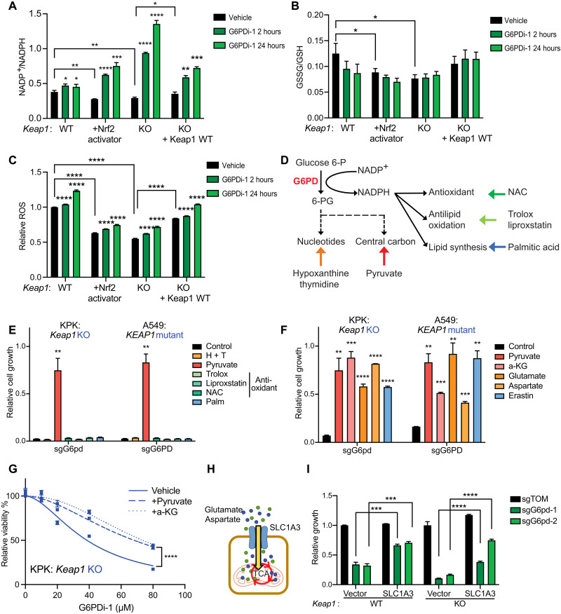
Fig. 2. G6PD loss or inhibition is rescued by central carbon metabolites but not antioxidants.
(A) NADP+/NADPH ratio of KP, KP + Nrf2 activator, KPK + vector, and KPK + Keap1 WT cells treated with 50 μM G6PDi-1 for 2 or 24 hours (n = 3), data measured by liquid chromatography–mass spectrometry (LC-MS). (B) GSSG/GSH ratio of KP, KP + Nrf2 activator, KPK + vector, and KPK + Keap1 WT cells treated with 50 μM G6PDi-1 for 2 or 24 hours (n = 3), data measured by LC-MS. (C) Relative ROS of KP, KP + Nrf2 activator, KPK + vector, and KPK + Keap1 WT cells treated with 50 μM G6PDi-1 2 or 24 hours (n = 3). (D) Schematic depicting G6PD biosynthesis pathway and corresponding rescue molecules. (E) Proliferation of KPK (Keap1 KO) and A549 (KEAP1 mutant) cells with G6PD KO in media supplemented with 30 mM hypoxanthine + 16 mM thymidine (H + T), 2 mM pyruvate, 50 μM trolox, 100 nM liproxstatin, 0.5 mM N-acetyl-l-cysteine (NAC), and 100 μM palmitic acid (Palm). Data were normalized to KPK sgTOM or A549 sgTOM (n = 3). (F) Proliferation of KPK (Keap1 KO) and A549 (KEAP1 mutant) cells with G6PD KO in media supplemented with 2 mM pyruvate, 2 mM dimethyl-2-oxoglutarate [a-ketoglutarate (a-KG) precursor], 6 mM glutamate, 20 mM aspartate, and 500 nM erastin. Data were normalized to KPK sgTOM or A549 sgTOM (n = 3). (G) Relative viability of KPK cells treated with G6PDi-1 and supplemented with 2 mM pyruvate and 2 mM a-KG for 3 days (n = 4). (H) Schematic depicting SLC1A3 up-taking extracellular glutamate and aspartate. (I) Proliferation of KP and KPK cells expressing SLC1A3 or an empty vector control with G6pd KO. Data were normalized to KP + vector with sgTOM group and KPK + vector with sgTOM group (n = 3). *P < 0.05, **P < 0.01, ***P < 0.001, and ****P < 0.0001.

