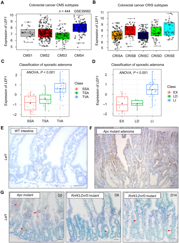
Fig. 1. LEF1 is expressed in Wnt ligand–independent but not in Wnt ligand–dependent CRCs.
(A and B) Analysis of LEF1 expression in the CRC (A) CMS and (B) CRIS subtypes. Data obtained from GSE39582. (C) Analysis of LEF1 expression (z score) in the sessile serrated adenoma (SSA), traditional serrated adenoma (TSA), and tubulovillous adenoma (TVA) CRC subtypes. (D) Analysis of LEF1 expression (z score) in the CRC subtypes. LI (ligand-independent tumor) has β-catenin mutation or APC mutation; LD (ligand-dependent tumor) has RSPO3 fusion or RNF43 mutation; and EX indicates samples without a known WNT alteration. (E and F) In situ hybridization of Lef1 (brown signal) in (E) adjacent normal tissue and (F) Apc-mutant adenoma. (G) In situ hybridization of Lef1 (brown signal) in Apcfl/fl;Villin-CreERT2 intestine 5 days after tamoxifen and in Rnf43fl/fl;Znrf3fl/fl;Villin-CreERT2 intestine 6 and 14 days after tamoxifen. Arrowheads point Lef1+ cells. Scale bars, 50 μm.

